LVH Criteria
- Amplitude of S wave in lead V1 + amplitude of R wave in V5 or V6 (whichever is the tallest) > 35 mm.
- R wave in aVL + S wave in V3 > 28mm men or 20mm
- R wave in aVL > 12mm
Understanding the Criteria
Left Ventricular Hypertrophy describes the increase in left ventricular mass that occurs as a response to chronic pressure or volume overload. The most common cause of pressure overload is systemic hypertension, but aortic stenosis as well.
As the size of the ventricle increases, the amplitude of QRS complex (which represents ventricular depolarization) also increases. Therefore, EKG manifestations of LVH are represented by large amplitude QRS complexes. The EKG leads that represent the left ventricle are V5, V6, I and AvL (see figure).
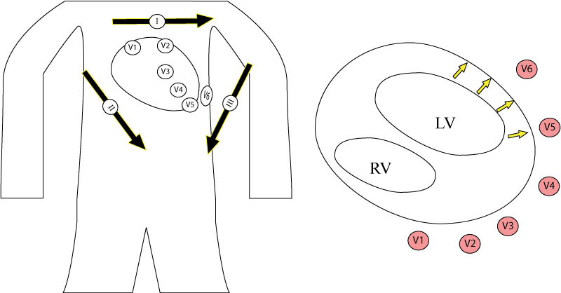
Remember that as a vector is larger towards a lead, the amplitude is more positive. If the vector moves away from a lead, then the amplitude is more negative. In the case of LVH, the increase manifests as large-amplitude R waves in lead I and AvL as well as the precordial leads closest to the left ventricle (V6) (see figure 2). By a similar logic, leads that are opposite the vector of the enlarged left ventricle have enlarged S waves (V5-V6).
There are many criteria for LVH, with varying sensitivity and specificity. The limb-lead criteria are more reliable in obese patients.
See examples of LVH here and here.