Mr P. is a 75 year old male admitted to the medical ward for a pneumonia. You are called by the nurse at midnight because the patient is feeling short of breath. Interpret his EKG.
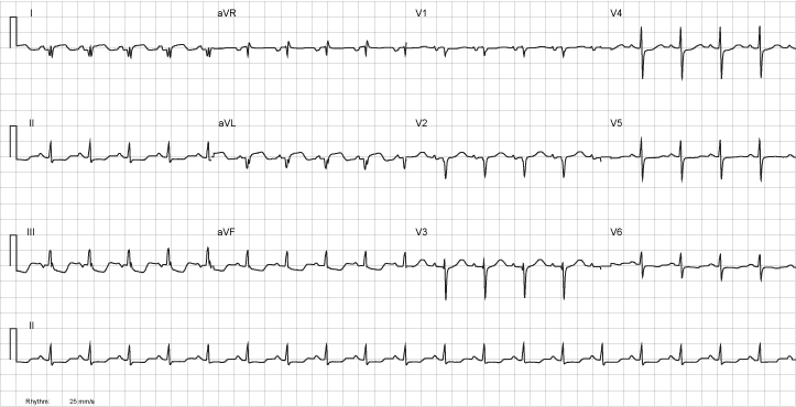
| Rate | 135 |
| Rhythm | Regular. P waves present. Sinus Tachycardia |
| Axis | RAD |
| Conduction |
|
| Hypertrophy | None |
| Infarction |
|
Discussion
This patient is having an acute myocardial infarction. The EKG is a an example of a lateral ST-elevation MI.
The ST elevations in the lateral leads (I and aVL) are indicative of acute myocardial injury. The presence of Q indicates the presence of infarction. In addition, seeing reciprocal ST- Depression in the inferior leads (II, III, aVF), further confirms the diagnosis. There is no evidence of anterior or septal involvement in the precordial leads.
Review the Infarction tutorial.