Mrs W. is a 77 year old female presents to the ER with dizziness. Interpret this EKG.
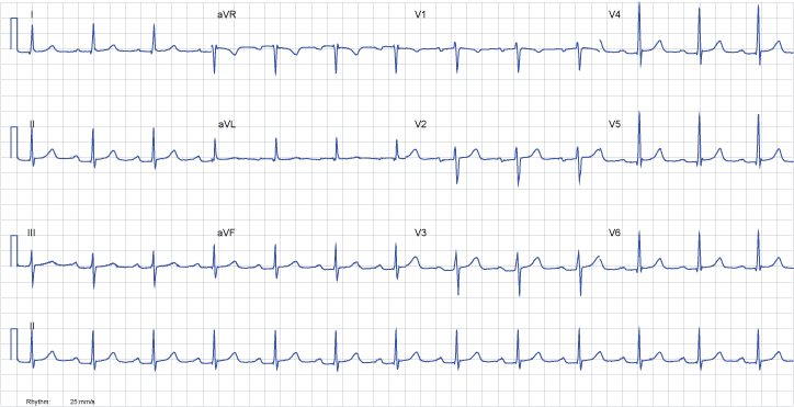
| Rate | 75 |
| Rhythm | Normal Sinus Rhythm |
| Axis | Normal |
| Conduction |
|
| Hypertrophy | None |
| Infarction | Non-significant q waves in I, aVL. Non-significant ST-T changes. |
This EKG is an example of a first degree heart block.
When you notice the elongated PR interval, you check that a QRS complex follows each p waves, and that the PR is of constant length in all of the complexes. If both of these are true, then you have a first degree heart block.
Could it explain her symptoms? Unlikely. Although it can progress to a higher degree block, a 1st degree heart block is an asymptomatic, benign condition.
The ST segments in this EKG seem suspiciously elevated, but the patient has some PR depression which exagerates the effect. Remember that the ST segment is measured in comparison to the TP segment.
Review the Rhythm tutorial .