Mr H. is a 34 year old female who presents for a pre-operative evaluation. What are the findings on her EKG?
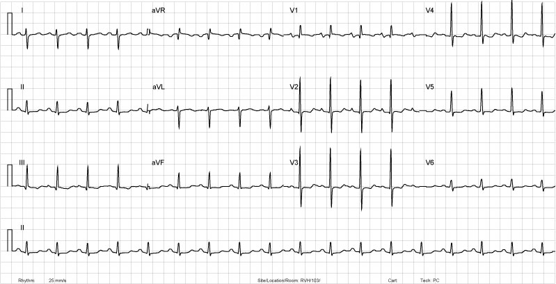
| Rate | 100 |
| Rhythm | Normal Sinus Rhythm |
| Axis | RAD |
| Conduction | QRS <0.12s |
| Hypertrophy | RVH |
| Infarction | insig. q waves III, aVF. |
Discussion
The patient has right ventricular hypertrophy .
In a normal EKG, the complexes in V1 and V2 should be negative. This is consistent with the diagnosis of RVH. The downward concavity of the ST segment and biphasic t waves is known as a RV strain pattern.
This would provide an explanation for the right-axis. Since the QRS is normal width, we can exclude a RBBB.
What would be a plausible explanation for RVH, LAE in a young 34 year old woman? What if on physical exam you detected an opening snap followed by diastolic rumble? Mitral Stenosis!
Review the RVH tutorial .
Review the Axis tutorial .