Mr H presents to the ER with heartburn. Interpret his EKG.
| Rate | 100-110 |
| Rhythm | Regular. P waves present. Sinus Tachycardia |
| Axis | Indeterminate |
| Conduction |
|
| Hypertrophy | No LVH or RVH |
| Infarction |
|
Discussion
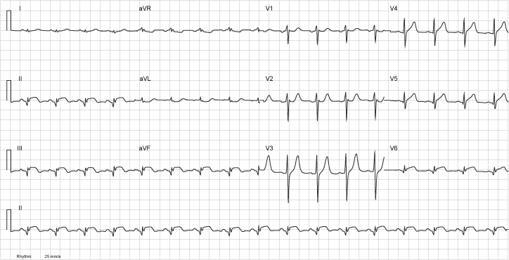
This EKG is very abormal. It has Q waves and ST elevation in the inferior leads (II, III, aVF). There is also involvement of the lateral leads labelling this as an inferolateral STEMI.
Leads I and aVL don`t show reciprocal changes, but these aren`t necessary for the diagnosis.
Review the Infarction tutorial .