You are seeing Ms. F in the Emergency Room. She is a 55 year old woman with a 30 pack year history of smoking who has presented with increasing cough and shortness of breath.
| Rate | 75-100 |
| Rhythm | Regular. P waves present. |
| Axis | Normal |
| Conduction |
|
| Hypertrophy |
|
| Infarction |
|
Discussion
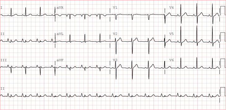
This patient has tall, peaked p waves in lead II. It is a classic EKG pattern called P Pulmonale associated with Right Heart strain. In a patient with a history of smoking, a COPD exacerbation is a likely cause. Remember to look at the shape of the p waves as part of a systematic interpretation. The ST changes in V2-3 are benign. On close inspection, the J point is well defined and doesn't appear to be elevated.