Mr L. is a 57 year old male with a known history of COPD, hypertension, and coronary artery disease. What are the findings on his EKG?
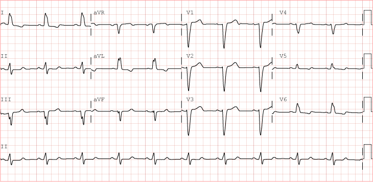
| Rate | 60 |
| Rhythm | Normal Sinus Rhythm |
| Axis | Normal |
| Conduction |
|
| Hypertrophy | None |
| Infarction | No Q waves. No ST-T abnormalities |
This EKG is a perfect example of a left bundle branch block.
Checking the width of the QRS complex should be part of a systematic EKG interpretation. When it`s longer than 120ms (3 small squares), then you should see if the pattern suggests a BBB morphology.
A LBBB can be a stable, chronic pattern, especially in patients with coronary artery disease. Interpreting ST elevations and hypertrophy is difficult with an LBBB. However, until proven otherwise, a new onset LBBB in the presence of chest pain is treated as equivalent to an ST-elevation MI. In this patient, correlating with an old EKG is essential .
Review the LBBB tutorial .