Among the most common hemetological tests performed are those which determine:
The first three tests (the red blood cell count, the hematocrit and the hemoglobin content) are used to calculate the Mean Corpuscular Volume (MCV) and the Mean Corpuscular Hemoglobin Concentration (MCHC).
Mammalian blood contains 5 different types of white blood cells which can be distinguished by staining with dyes. In this test you will be determining the percentages of each type of leukocyte present in the blood.
Differential White Blood Cell countHow to differentiate the five types of the WBC.
|
| Granulocytes: |
Neutrophils, eosinophils and basophils are described collectively as granulocytes. They are distinguished by the nature of the granules in their cytoplasm, and generally have small, multilobed nuclei. |
| Image of the WBC |
Description |
Colors of the cell parts |
Expected Renges |
 |
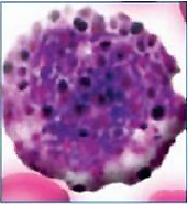
Neutrophils have nuclei with several lobes and fine purple granules in their cytoplasm. Their are termed neutrophil, because their granules are not very amenable to staining with either acidic or basic dyes. They are the most numerous of the leukocytes. |
Polymorphonuclear neutrophils:
nucleus: dark blue
cytoplasm: pale pink
granules: reddish lilac |
50-70% |
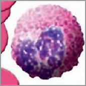 |
Eosinophils feature large granules that can be stained bright red with an acidic dye - e.g. eosin. They most often have bi-lobed nuclei. |
nucleus: blue
cytoplasm: blue
granules: red-orange |
1-4% |
 |
Basophils have multi-lobulated nuclei and large granules which stain blue with basic dyes. They occur quite seldomly. |
nucleus: purple or dark blue
granules: dark purple, almost black |
0.1% |
| Agranulocytes |
The 2 other types of white cells are lymphocytes and monocytes and are described collectively as agranulocytes. |
| Image of the WBC |
Description |
Colors of the cell parts |
Expected Renges |
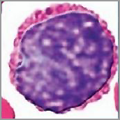 |
Lymphocytes can be large or small. They are spherical and have a very large nucleus taking up most of the cytoplasm. The cytoplasm has no granules. |
nucleus: violet
cytoplasm: dark violet
|
20-40% |
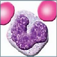 |
Monocytes are large cells. They have large indented nuclei, often kidney-shaped and very fine cytoplasmic granules. |
nucleus (lobated): violet
cytoplasm: sky blue |
2-8% |
|
Procedure:
Place the prepared slide on the microscope and scan with the low power to find an area with a good distribution of cells. Place a drop of oil on the slide and examine it with the oil immersion objective (x10
| First, practice identifying each cell type. Then count a total of 100 white blood cells and record the number of each type seen (e.g. 2 eosinophils, 25 lymphocytes, etc.) |
| Neutrophils |
Eosinophils |
Basophils |
Lymphocytes |
Monocytes |
|
|
|
|
|
| |
|
|
|
|
|
Add the total number of each type of WBC and determine the grand total. Use this number to calculate the % of each type of WBC and enter these values in the table in your lab report. Multiply the % by the value you obtained for the total WBC count to calculate the number per mm3 of each type of WBC, and add these values to the table in your lab report.
Practice
If you want to practice before your class, click here to open a window which mimics
what you might see looking through the 100x objective of the microscope
using oil immersion. Try to determine the differential white cell count.
|
| Leucocute types |
Percent (%) |
# of cell / mm3 (% x Total WBC count) |
| Neutrophils |
|
|
| Eosinophils |
|
|
| Basophils |
|
|
| Lymphocytes |
|
|
| Monocytes |
|
|
|