PHGY 212    Blood Lab
Blood Cell Indices

Among the most common hemetological tests performed are those which determine:

The first three tests (the red blood cell count, the hematocrit and the hemoglobin content) are used to calculate the Mean Corpuscular Volume (MCV) and the Mean Corpuscular Hemoglobin Concentration (MCHC).

Red and Total White Blood Cell count

Procedure


This video shows hemocytometer loading.

However, there are differences (listed below) between Red and White Blood Cell count.

Differences between Red and White Blood Cell count preparation

  RBC WBC
dilution reagent Gower's solution glacial acetic acid
dilution 1:200 1:20

Red and White Blood Cell counting


RBC    

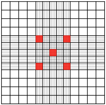
Hemocytometer grids contain 9 large, 1 mm x 1 mm squares. RBCs are counted in the central square of the hemocytometer. Note that the central square is divided into 25 smaller, 0.20 mm x 0.20 mm squares.
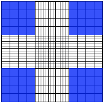
In this lab, you will count five squares out of the 25 squares within the central grid (see on the left).

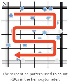
If we zoom in on one of the five squares you numbered above, you will see that it is further divided into 16 squares. To count RBCs, scan these squares from top to bottom in a serpentine pattern, as shown on the right.

Follow these rules when counting RBCs inside squares that touch an outside boundary (indicated by three parallel lines):
- Include any cells that touch the middle line of the top or left border, even if part of the cell touches the outside boundary of the square.
- Exclude any cells that touch the middle line of the bottom or right border.

WBC    

Hemocytometer grids contain 9 large, 1mm x 1mm squares. WBCs are counted in the four 1mm x 1mm corner squares of the hemocytometer grid which are also subdivided in 16 smaller aquares.

Similar to counting RBCs, if we zoom, in this case, on one of the four corner squares (see on the left), you will see that it is further divided into 16 squares. To count WBCs, scan these squares from top to bottom in a serpentine pattern, as shown above.

On the right you can see the top left corner square of the Hemocytometer grid. Notice different shapes.

  • A - example of debris (big dark particles)
  • B - example of a WBC (they look like small black dots)
  • C - examples of light gray debris - membranes of broken down RBCs

Microscope

There are 3 lenses: 10x, 40x and 100x. The first - 10x, is used to locate the area of interest. Then we switch to the lens appropriate for the specimen. For the RBC and Total WBC count we use 40x. The 100x lens requires a use of the immersion oil. It is used for the Differential WBC count.

Red and White Blood Cell count calculation

  RBC WBC
formula (cells/mm3) 200 x 10 x 5 20 x 10 / 4