PHGY 212    Immunology Lab
Hemagglutination


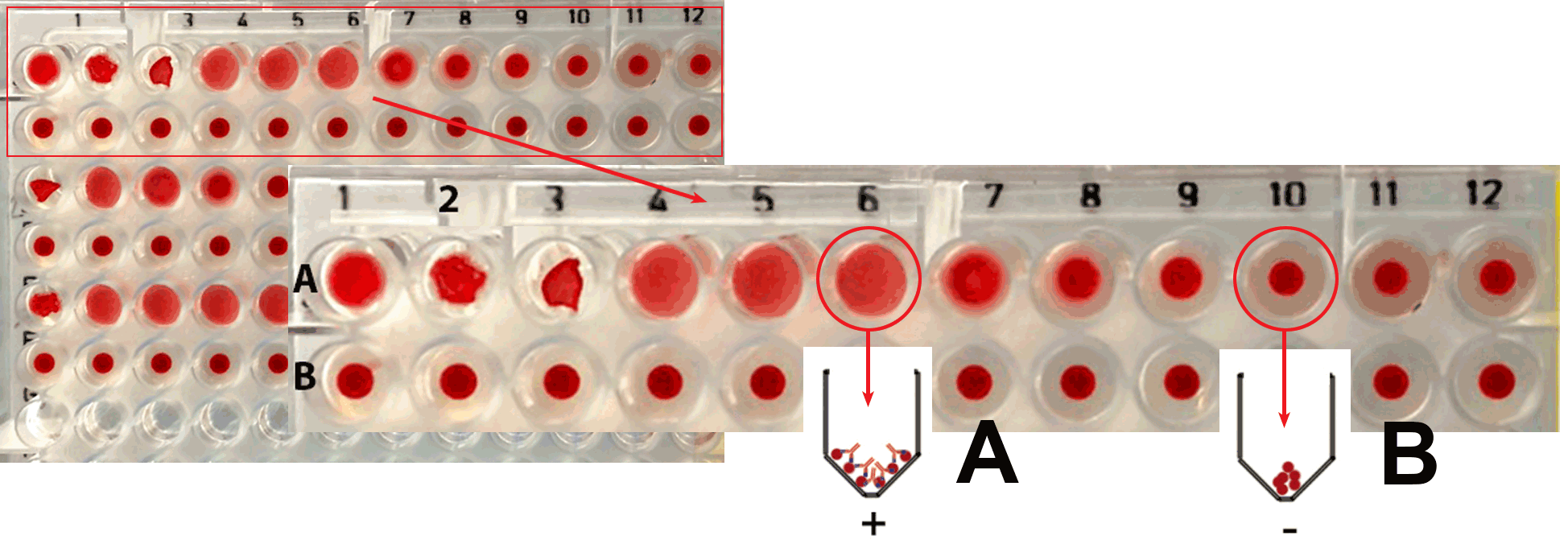
Antibodies can be detected in the serum of animals. If red blood cells are used as a source of antigen, the assay is called hemagglutination. An animal injected with Sheep red blood cells (SRBC) will develop antibodies against SRBC. In the experiment, SRBC are added to serially diluted serum from an immunized animal. The antibodies in the serum will agglutinate the SRBCs added in a test well, resulting in a specific pattern at the bottom of the well.


Hemagglutination:


Mandatory revision of serial dilutions

Procedure:

Hemagglutination (Video transcript)

Agglutination is a technique for determining that specific antibodies and their antigens are present in a reaction. It is also used to measure the titer, the concentration, of an antibody.

Hemagglutinaton was once widely used for blood typing preceding blood transfusion. But blood typing is now done using other methods. Agglutination techniques are still used to evaluate bacterial diseases like cholera.

In this type of experiment, antigens and serum containing antibodies are mixed together. If a specific match is present, there will be cross linking of the two, due to the bivalent nature of the antibodies. This results in the formation of a precipitate or an aggregation or agglutination reaction that can be measured. (This concept will be revisited in the blood lab)

With the agglutination technique, the antigen is often a surface protein on a cell, like a red blood cell or a bacterial cell.

In our experiment, the antigen is a protein on the surface of sheep red blood cells and we will use serum from two patients as a source of antibodies. When we mix the RBCs with a serum that contains antibodies specific for surface antigens on the RBC, we will have a positive interaction that results in the agglutination of the RBCs. This is called hemagglutination.

To determine the antibody titer (concentration), we will dilute the serum and perform the reaction over a range of serum concentrations. Hemagglutination is maximal when there is an equal proportion of antibody and antigen. Our goal is to find the dilution of serum that produces maximal hemagglutination. This will be the antibody titer.

Our hemagglutination experiments will be conducted using 96-well microtiter plates. The plate contains 12 wells in each of its 8 rows.

We will measure the titers of two serum samples: Serum 1 and Serum 2. Other reagents we will use in this experiment are normal saline and sheep red blood cells, which will serve a source of antigen.

In Row A we will test serum sample 1. In Row B we will test serum sample 2 and the first four wells of Row C will contain our negative controls which will not have any serum added.
  • STEP 1: We will begin by placing 100 microliters of saline in all well of rows A and B, and in the first four wells of Row C.
  • STEP 2: Place 100 microliters of Serum 1 into the first well of row A and proceed with a 2-fold serial dilution. A 2-fold serial dilution involves mixing the serum added to the first well with the equal volume saline already in it by carefully pipetting up and down. 100 microliters of this mixture is then moved to the second well in the same row. We mix again and transfer 100 microliters of this mixture to the third well in the row. We continue this procedure for all the wells in the row. Using this technique, the concentration of serum found in the original tube will be halved in each well we move across the plate from left to right. When we reach well twelve, we will mix the contents and remove and discard the additional 100 microliters
  • STEP 3: Place 100 microliters of Serum 2 in the first well of row B.
  • STEP 4: Perform the two-fold serial dilution for row B, again discarding the extra 100 microliters in well twelve. Be sure to use a separate pipette tip for each row so that we do not cross contaminate our samples.
  • STEP 5: We will now add 100 microliters of sheep red blood cells to all the wells of row A and B and the first four wells of Row C. It is important to mix the tube of red blood cells before using it to ensure that we have a homogeneous mixture.
  • STEP 6: When pipetting the sheep red blood cells we can use the same pipette tip for all the wells if we are careful to avoid touching the liquid in each well. There is no need to mix the contents of the well with the RBCs.
  • STEP 7: Cover the plate with an adhesive plastic film and press to adhere. Label your plate.
  • NOTE: It is important in this and all subsequent steps, to not invert your plate.
  • STEP 8: Incubate the plate at 37-degree C for 30 minutes.
  • STEP 9: Move the plate to the refrigerator at 4 degrees C for several hours or overnight, as time permits.
  • STEP 10: Read the results of your plate by looking at it from the bottom (without inverting the plate) to evaluate the amount of hemagglutination in each well.

Results:

Antibody may be detected and measured by hemagglutination at lower concentrations than those detectable by other techniques. This relies on the ability of antibody to cross-link red blood cells by interacting with the antigens on their surface. 
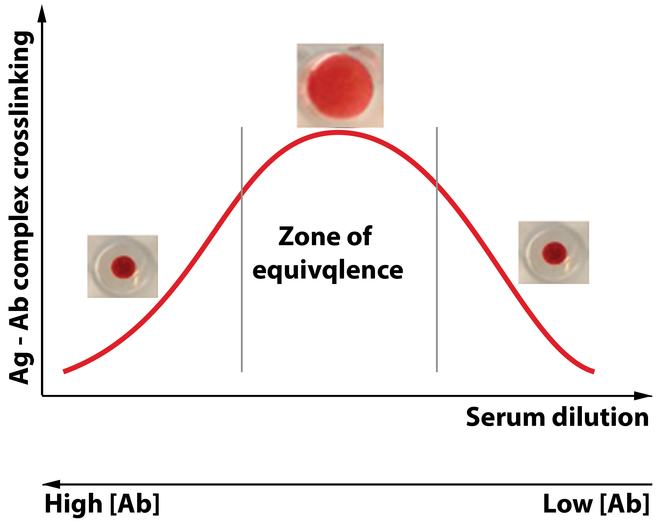
The agglutination of an antigen, as a result of crosslinking by antibodies, is dependent on the correct proportion of antigen to antibody.

Reading the 96-well plate:

A. If sufficient antibody is present to agglutinate and form cross-linking with the antigen, the antibody-antigen complex forms a mat at the bottom of the well.

B. If insufficient antibody is present, the cells roll down the sloping sides of the well to form a red pellet or "button" at the bottom of the well.

Discussion:

In the zone of equivalence, the correct proportion of antibody to antigen occurs, resulting in a visible mat formed by Ag-Ab complex crosslinking.

At high concentration of antibodies: Ag-Ab complex crosslinking is prevented because every epitope on an antigen particle may bind to a single antibody molecule. At higher dilutions, agglutination may occur: crosslinking is possible.

Testing serum at only one concentration may give misleading conclusions. What might the absence of agglutination reflect?