PHGY 212    Immunology Lab
Complement Mediated Lysis


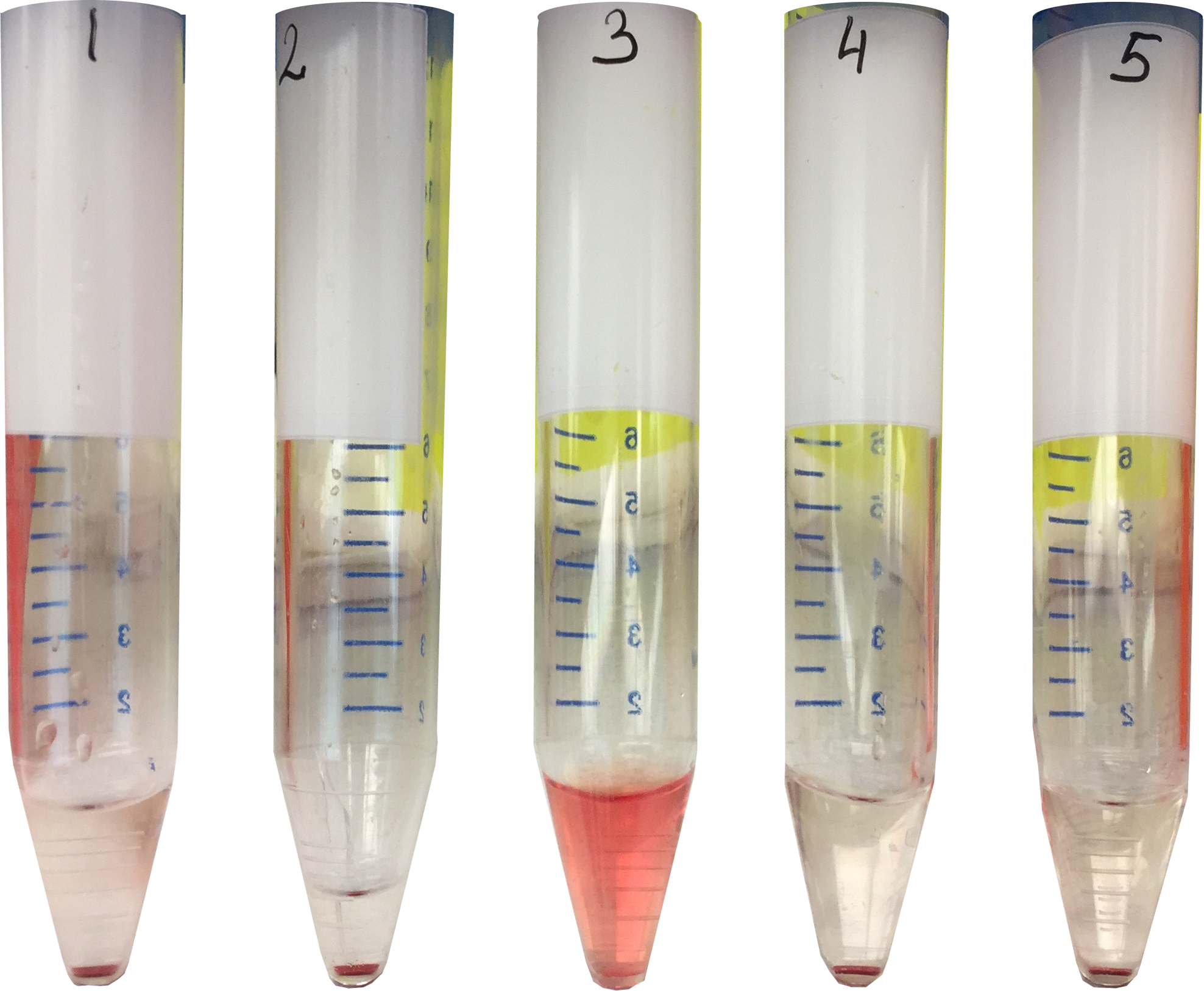
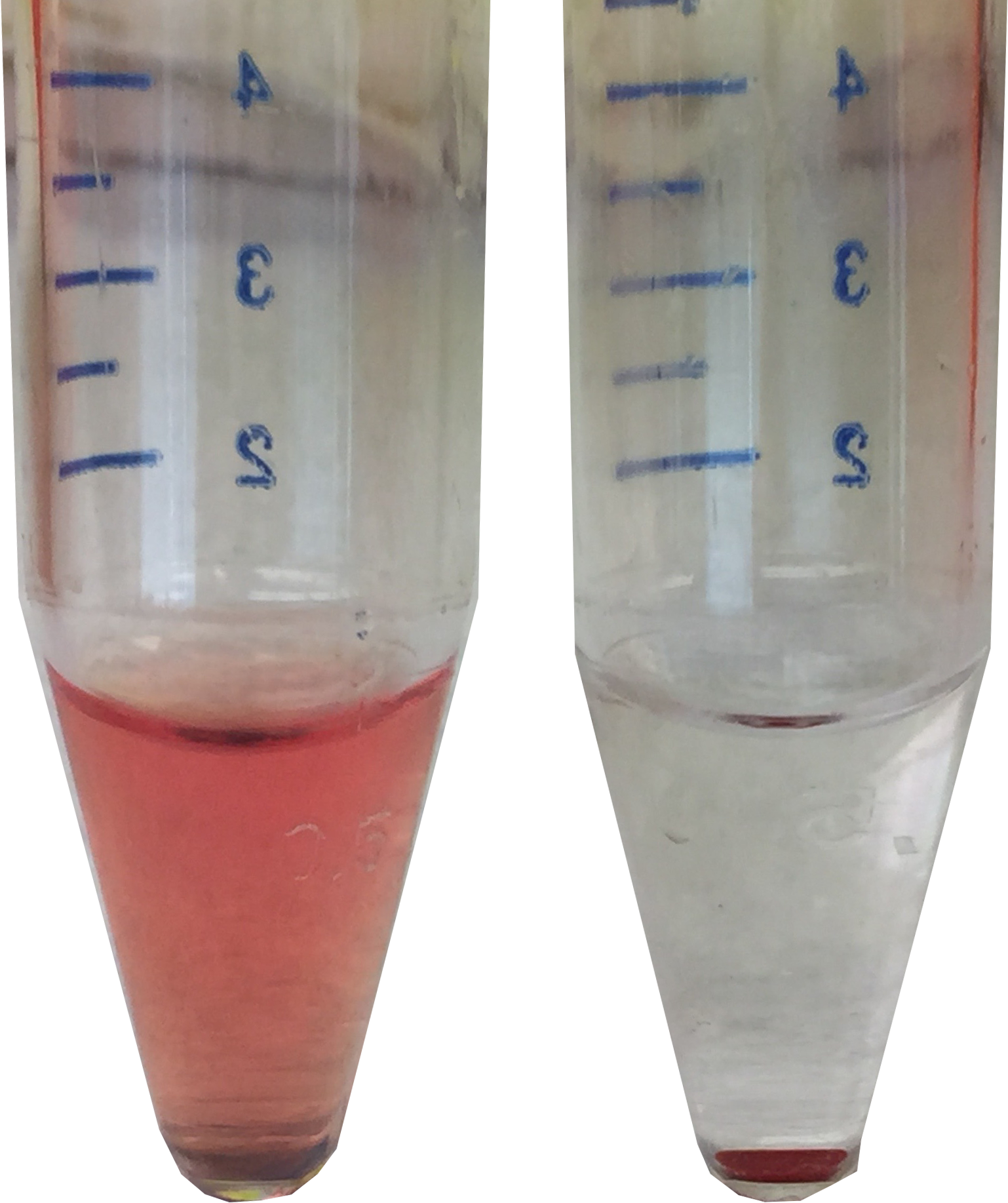
Antibody by itself is rather ineffectual in eliminating foreign organisms. However, antibody of the IgM and IgG class can activate the Complement (C') system resulting in a stimulation of different effector functions such as phagocytosis and lysis of foreign organisms.

In our experiment Sheep Red Blood Cells (SRBC) are used as a source of antigen to demonstrate the lytic properties of the antigen-antibody-complement complex

Procedure

Complement Mediated Lysis / Cytotoxicity (Video transcript)

The complement system plays several important roles in immunity. The complement system can cause the lysis of bacterial cells through the formation of a Membrane Attack Complex which makes holes in the targeted cell, causing its death.

This experiment demonstrates the importance of the complement system in immune protection and allows us to determine the sequence of events necessary for this process to occur.

We will conduct these experiments in 15 ml conical tubes.

First we will label the tubes from 1 to 5. Each tube will contain a different mixture of components. Which will allow us to determine the sequence of events needed for complement mediated lysis to occur.

We will now add the components to each tube, according to the table found in our lab protocol.

We will begin with the sheep Red Blood Cells. We make sure that the RBCs are well-mixed – so that we have a homogenous suspension of cells.

We will begin by adding 300 microliters of well-mixed sRBC to each tube.

In this experiment sheep Red Blood Cells are the source of antigen. Formation of the Membrane Attack complex on their surfaces, will cause the lysis of the RBCs.

In tubes 2 and 3 we will add 300 microliters of antiserum. This antiserum comes from a rabbit that was immunized with an antigen specific to the surface proteins of the sheep RBCs.

In tube 4 we will add 300 microliters of normal rabbit serum. Normal rabbit serum contains antibodies, but none that are specific to the RBC surface antigen.

We will now add 300 microliters of saline to tubes 2 and 5.

We will add 300 microliters of complement to tubes 3, 4 and 5. This complement was obtained from guinea pig serum. It contains purified complement molecules.

Finally we will add 600 microliters of saline to tube 1.

Now that we have added all the components to the tubes we will allow the components to incubate. This will allow time for the membrane attack complex to form.

We place the tubes in a rack in a water bath set at 37 degrees C and incubate for 30 minutes.

Based on what you know about the components in each tube, make a prediction about which tubes will undergo red blood cell lysis.

After 30 minutes has elapsed, we will remove the tubes from the water bath, and note any differences in the appearance of the contents.

The contents of one tube appear as a relatively clear pink solution, while the others have a more cloudy pink colour and some of the intact red blood cells have settled to the bottom of the tubes.

We will centrifuge our tubes at maximal speed for 5 minutes, being sure that our tubes are properly loaded and balanced.

After five minutes, we remove the tubes and note any differences in appearance of the contents. In the tubes in which the RBCs have not lysed we see a red pellet containing the intact RBCs and a clear supernatant. in the tube where lysis occurred the solution pink.

Record the results in our lab notebook.

We will continue the experiment with tubes 2 and 4 in which a pellet has formed. We begin by dumping the supernatant into a waste container. We will wash the pellet by adding 3 milliliters of saline and then gently resuspending the pellet using a Pasteur pipette. Washing the pellet will remove any reagents that are present in the supernatant and that are not bound to the surface of the red blood cells.

One we have added the 3 ml of saline to both tubes we will gently resuspend the pellet using a Pasteur pipet. it is important that we use different Pasteur pipettes for each tube to avoid cross contamination of our samples.

We will centrifuge the tubes for 5 minutes at maximal speed and then dump off the supernatant.

According to the TABLE we will add the following reagents:
- To both tubes we will add 300 microliters of saline.
- We will add 300 microliters of complement to tube 2.
- We will add 300 microliters of antiserum to tube 4.

We will now gently re-suspend the pellets with a Pasteur pipette. Again we will use a clean Pasteur pipette to resuspend each pellet to avoid cross contamination.

With our components added we can place the two tubes in a rack in a water bath set at 37 degrees C and allow them to incubate for 30 minutes.

Use this time to predict your results.

Note the difference in the appearance of the contents even before centrifugation.

We will centrifuge the tubes for 5 minutes at maximal speed.

We can now record our results in our laboratory notebook.

Results

Part 1

Upon centrifugation the red blood cells in the tubes where cells have undergone lysis, the solution will appear pink (tube 3)

In the tubes where no lysis occured, the solution will appear clear and the intact red blood cells will be found in a pellet at the bottom of the tube. (tubes 1,2,4,5)

Part 2

Upon centrifugation the red blood cells in the tubes where cells have undergone lysis, the solution will appear pink (tube 2)

In the tubes where no lysis occured, the solution will appear clear and the intact red blood cells will be found in a pellet at the bottom of the tube. (tube 4)