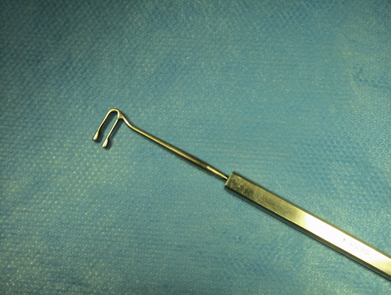
Du Plessis Muscle Hook
Similar in design to the Jameson hook, the Du Plessis has 2 hooks instead of 1. The additional hook elevates the rectus muscle away from the globe and provides additional protection when weaving the suture through the muscle.
Du Plessis muscle hook MR
This video demonstrates the use of the Du Plessis muscle hook for the exposure, suturing and disinsertion of the medial rectus muscle. The left eye is positioned with the medial rectus to the right and the inferior rectus at the top of the screen. A limbal conjunctival incision has been completed nasally and inferiorly. The medial rectus insertion is engaged with a Jamieson hook superiorly and then inferiorly. After it is freed of check ligaments and intermuscular membranes, a Du Plessis hook is inserted under the medial rectus and just behind the insertion. The Jamieson hook is removed. A 6-0 double-armed vicryl suture is woven through the muscle near its insertion site. The Du Plessis hook facilitates this maneuver by elevating the muscle above the scleral plane and by providing a safe channel between the double hooks. Inferior and then superior fixation of the muscle with the suture is completed. The hook is then exchanged for a Jamieson hook and the muscle is disinserted. The 2 arms of the suture are reinserted at the insertion site and the muscle allowed to hang back a measured distance from the insertion site. The 2 arms of the suture are tied in a bow to be used for future adjustment purposes.