|
Immunology Laboratory |
Hemagglutination |
| |
Antibody can be detected in
the serum of animals. If red blood cells are used as a source of
antigen, the assay is called hemagglutination. An animal receiving Sheep
red blood cells (SRBC) will develop antibodies against SRBC. In
the experiment, SRBC are added to serially
diluted serum from this immunized animal. The antibodies in the serum will
agglutinate to SRBC added in a test tube, resulting in a specific
looking pattern at the bottom of the tube. Hemagglutination
|
|
Procedure |
|
You are given two sera, from a "normal"
mouse and one that was immunized with SRBC.
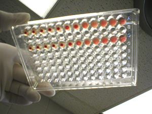
Using micro titer plates place 0.1 ml of
balanced salt solution into each well of rows A and B. Add 0.1 ml of
serum #1 to the first well, mix and transfer 0.1 ml to the next well in
row A and so on until you reach the end.
Repeat the procedure for serum 2 in row
B. Add 0.1 ml of a 1% suspension of SRBC. Incubate for 30 minutes at 37C
and then place in the refrigerator. Best results are obtained if you
examine the plates the next morning; however you should have results by
the end of the laboratory session. Explain your results and report the
titer of the two antisera. |
Serial
dilution (doubling dilution): this technique is referred to
as serial dilution, where you dilute a reagent or compound in series or
in sequence. Since you take the same quantity of reagent from one well
to the other, and the first well has the same volume as you put in: you
are doubling your dilutions.
|
|

|
|
Results |
 |
Antibody may be detected and measured by
hemagglutination at lower concentrations than those detectable by other
techniques. This relies on the ability of antibody to cross-link red
blood cells by interacting with the antigens on their surface.
The agglutination of an antigen, as a result of crosslinking by
antibodies, is dependent on the correct
proportion of antigen to antibody. |
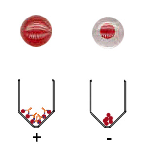
Reading the 96-well plate:
If sufficient antibody is present to agglutinate and form
cross-linking with the antigen, the antibody-antigen complex forms a
mat at the bottom of the well. If insufficient antibody is present, the
cells roll down the sloping sides of the well to form a red pellet or
"button" at the bottom of the well. |
 |
Hemagglutination is expressed as
titer: it is the inverse of the last
dilution that is positive; e.g: 1/1000. |
|
Discussion |
|
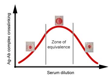
In the zone of equivalence, the correct
proportion of antibody to antigen occurs, resulting in a visible mat
formed by Ag-Ab complex crosslinking.
At high concentration of antibodies: Ag-Ab complex crosslinking is
prevented to occur; every epitope on one antigen particle may bind to a
single antibody molecule.
At higher dilution of serum, agglutination may occur: crosslinking is
possible. Why?
Testing serum at only one concentration may give misleading conclusions.
What might the absence of agglutination reflect? |
 |
|
Depending on the starting
dilution, the figure above could be derived. |
|
|
|
Click here to
continue with the topic of Complement cytotoxicity |