|
Immunology Laboratory |
Complement mediated
cytotoxicity |
| |
Complement mediated
cytotoxicity is an effector function assay. Antibody by itself is rather
ineffectual in eliminating foreign organisms. However, antibody of the
IgM and IgG class can activate the Complement (C') system resulting in a
stimulation of different effector functions such as phagocytosis and
lysis of foreign organisms.
|
|
|
If red blood cells are used as a source of
antigen it is relatively simple to demonstrate the lytic properties of
the antigen-antibody-complement complex. |
|
Procedure |
|
Take 5
tubes and label them 1 to 5. In each tube place 0.3 ml of SRBC (1%
solution). In tubes 2 and 3 place 0.3 ml of antiserum and in tube 4
place 0.3 ml of normal mouse serum. In tube 1 place 0.6 ml of saline and
tubes 2 and 5 place 0.3 ml of saline. In tubes 3, 4 and 5 add 0.3 ml of
complement solution. Place in the water bath at 37C and incubate for 30
minutes. Remove from the water bath and centrifuge. Record the colour of
the solution in each tube and explain the reason for the differences
observed. |
|
Tube: |
1 |
2 |
3 |
4 |
5 |
|
Add: |
 0.3
ml SRBC (1%) 0.3
ml SRBC (1%) |
|
|
0.3ml anti-serum |
0.3ml anti-serum |
0.3ml normal mouse serum |
|
|
0.6ml saline |
0.3ml saline |
|
|
0.3ml saline |
|
|
|
|
0.3ml C’ |
0.3ml C’ |
0.3ml C’ |
|
Incubate: |
In water bath for 30 minutes |
|
Centrifuge: |
|
|
Record: |
Colour change |
|
After recording your results, take tubes 2
and 4 and add 3 ml of saline to each tube and gently resuspend the RBC
pellet with a Pasteur pipette. Centrifuge them gently for 5 minutes to
form a RBC pellet. Remove the tubes from the centrifuge and aspirate off
the supernatant with a Pasteur pipette.
To each tube add 0.3 ml of saline, and to tube 2 add 0.3 ml of C'
solution and to tube 4 add 0.3 ml of antiserum. Mix gently with a
Pasteur pipette and incubate at 37C (water bath) for 30 minutes,
centrifuge for 5 minutes. Record the colour change and interpret your
results.
What do you conclude about the specificity of antibody and complement
binding? |
|
Tube: |
2 |
4 |
|
Add: |
0.3
ml saline (after washing the cells, centrifuging, removing the
supernatant) |
|
0.3ml C' |
0.3ml antiserum
|
|
Mix gently with Pasteur
pipette |
 |
|
Incubate: |
In water bath for 30 minutes |
|
Centrifuge: |
5 minutes |
|
Record: |
Colour change |
|
|
Results |
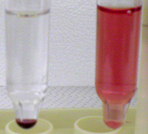 |
Upon centrifugation, the cells "made up" in
clear saline will collect into a pellet at the bottom of the
tube and since no lysis has occurred, there will be a clear saline
supernatant over the pellet (left tube).
If lysis has occurred, the release of sheep red blood cell contents in
the supernatant will occur and a "ghost" pellet made up of cell membrane
debris will be at the bottom of the tube (right). Which sequence of events caused
cell lysis? |
Click here to return to the virtual lab home page |