|
Blood
Laboratory |
Blood cell indices
>
Differential white cell
count > Practice |
| |
When the blood smear has dried and been stained with
Diffquick, the slide is placed on a microscope and scanned at low power to find a good
distribution of cells. A drop of oil is placed on the slide and the cells are examined
with the oil immersion objective. The percentage of each type of white blood cell is
determined. |
|
|
|
Preparation of the slide:
(procedure not done during the laboratory session; however,
pre-stained slides will be available for leukocytes differential count) |
|
|
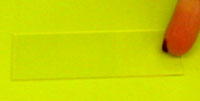
1) A fresh (non-heparinized) sample of blood
is added to one side of the slide |
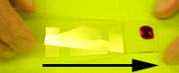
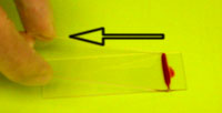
2) The edge of another slide is pushed
against the drop of blood and smeared onto the rest of the slide (see 3
and 4 below). |
|
3) |
4) |
|
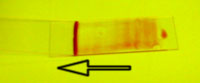
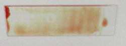 The
smeared slide is allowed to dry. The
smeared slide is allowed to dry. |
|
The staining procedure: |
|
 |
|
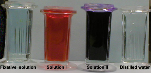
Diff-Quik Stain set is a
modification of the Wright Stain technique:
Blood smears are fixed using the methanolic fixative solution in order
to stabilize cellular components. Solutions I and II are then applied
individually to the fixed smear to differentially stain specific
cellular components. |
|
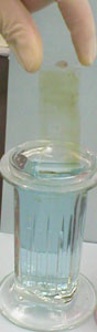
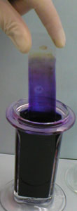
The dried slide is dipped
several times in the Fixative solution. The excess is allowed to
drain. |
|
 |
|
 |
|
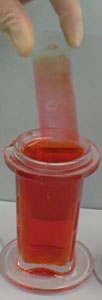
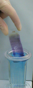
Then this slide is dipped several times in
Solution I, which is a buffered solution of Xanthene dye (an anionic
dye). The dye stains the granules in the cytoplasm, a bright orange
colour. |
|
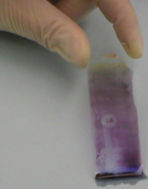
The same slide is dipped several times in
Solution II, which is a buffered solution of thiazine dyes (cationic
dyes) consisting of methylene blue and Azure A.
The resultant basophilic staining of nucleoli and cytoplasm is due to
the methylene blue component of the mixture. The anionic component of
the nucleoli and cytoplasm is stained with the cationic methylene blue. |
|
 |
 |
| The slide is rinsed with
distilled water and allowed to dry.
|
|
| Leukocytes: |
| |
Granular |
| |
|
Polymorphonuclear neutrophils |
| |
|
nucleus: dark blue |
| |
|
cytoplasm: pale pink |
| |
|
granules: reddish
lilac |
| |
|
Eosinophils |
| |
|
nucleus: blue |
| |
|
cytoplasm: blue |
| |
|
granules: red-orange |
| |
|
Basophils |
| |
|
nucleus: purple or
dark blue |
| |
|
granules: dark
purple, almost black |
| |
Non-granular monocytes |
| |
|
nucleus (lobated):
violet |
| |
|
cytoplasm: sky blue |
| |
Lymphocytes |
| |
|
nucleus: violet |
| |
|
cytoplasm: dark blue |
|
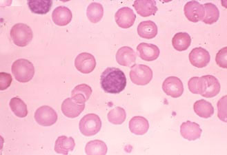
The slide is examined under oil immersion. |
|
 |
| Expected Ranges |
| Neutrophil (%) |
50-70 |
| Eosinophil (%) |
1-4 |
| Basophil (%) |
0.1 |
| Monocyte (%) |
2-8 |
| Lymphocyte (%) |
20-40 |
|
|
Click here to open a window which mimics
what you might see looking through the 100x objective of the microscope
using oil immersion. Try to determine the differential white cell count. |
|
To continue
with the next section, blood typing, click here |