|
Compound Action Potential |
Background >
Nerve Anatomy |
|
A typical nerve in a
vertebrate like the frog consists of several
thousand axons. For example, the vagus nerve in man consists of
over 100,000 fibers.
The cell bodies of these axons are located either in the central nervous
system (for motor fibres), or in the peripheral nervous system (e.g.
dorsal root ganglia, for sensory fibres).
The fibres may be large (15-25 µm) and myelinated, or small (around 0.2
µm) and unmyelinated. Regardless of the type or size of the fibres, they
all have certain properties in common; e.g., they
all conduct non-decremental action potentials.
However, there are also certain differences,
e.g. the conduction velocity of the fibres
differs, depending on their size (diameter)
and whether they are myelinated or non-myelinated. |
|
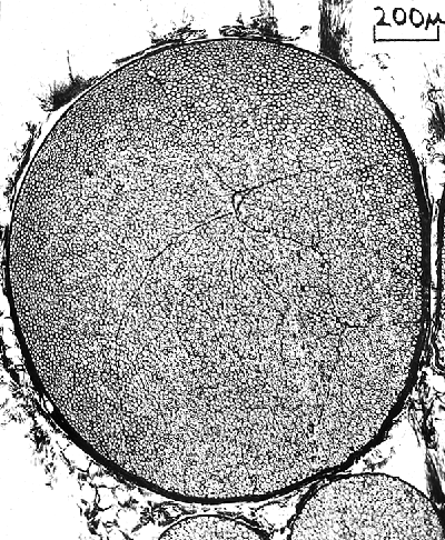
Cross-section through an unmyelinated nerve. |
|
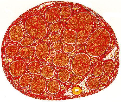
Cross-section through a myelinated nerve
(e.g. sciatic) showing individual nerve bundles each consisting of many
fibres. |
|
|
In general, small (less than 25mm),
myelinated and unmyelinated fibres are a feature of vertebrates, while
large (up to 500 mm
or larger), unmyelinated fibres are a feature of invertebrates (e.g. the
squid) |
|
In vertebrates (including man), the whole
nerve is encased in a connective tissue sheath called the Epineurium.
The axons are further subgrouped into bundles called fasciculi
(singular: fasciculus), each of which is also encased in a connective
tissue sheath called the Perineurium. Lastly, each individual fibre is
further surrounded by a delicate connective tissue sheath called the
Endoneurium. |
|
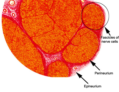
A higher
magnification of several nerve fibre bundles showing individual axons as
distinct circular structures.
|
|
|
|
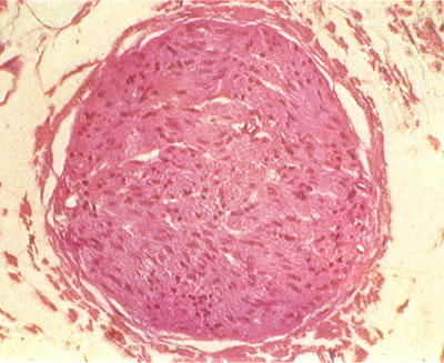
The frog's sciatic consists of only a single
bundle of fibres, surrounded by the perineum and loose epineurium |