During pregnancy, normal
trophoblast cells of the placenta secrete a glycoprotein hormone: human
chorionic gonadotropin (hCG). Because, under normal circumstances, hCG
is produced only by placental tissue and is conveniently found in the
blood or urine of a pregnant woman, hCG detection is a sensitive
chemical test of early pregnancy.
Early development, implantation and
placentation
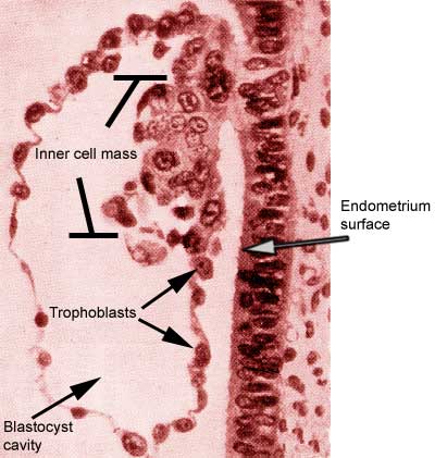
After reaching the uterus, the fertilized
egg or zygote - getting nutrients from the intrauterine fluid -
undergoes further cell divisions and reaches the stage known as
blastocyst whereby the cells begin to differentiate. The blastocyst
consists of an outer layer of cells, the trophoblast, an inner cell mass
and a central fluid-filled cavity. The inner cell mass will give rise to
the embryo and some of the membrane associated with it. The trophoblast
will surround the embryo throughout development and be involved in its
nutrition as well as in the secretion of several important hormones.
Blastocyst
implanted in the endometrium
The zygote-to-blastocyst
development period corresponds with days 14 to 21 of the typical
menstrual cycle. At this time, the uterine lining is being prepared by
progesterone secreted by the corpus luteum to receive the blastocyst. By
the 21st day of the cycle or 7 days after ovulation, the blastocyst
embeds itself into the endometrium. The trophoblast cells are very
sticky especially in the region around the inner cell mass. It is this
portion of the blastocyst which adheres to the endometrium and initiates
implantation. Rapid trophoblast proliferation occurs and cells penetrate
between endometrial cells.
The nutrient-rich endometrial cells provide what is necessary for early
growth of the embryo, but this function is replaced after the first few
weeks by the placenta: a combination of interdigitating fetal and
maternal tissues. The fetal portion of the placenta is supplied by the
outermost layers of trophoblast cells: the chorion. Fingerlike
projections of the trophoblast cells (chorionic villi) extend from
the chorion into the endometrium. Human chorionic gonadotropin (hCG)
starts to be secreted by the trophoblast cells around the time they
start their endometrial invasion. The hCG enters the maternal
circulation and thus can be detected.
Fetal
and maternal tissues
Function of the human chorionic gonadotropin: without
pregnancy, the corpus luteum degenerates within two weeks after its
formation. The function of HCG is to maintain the corpus luteum. This
hormone also strongly stimulates steroid secretion by the corpus luteum
in the beginning stages of gestation.
Hormonal changes during pregnancy
The secretion of hCG reaches a peak 60 to 80
days after the last menstrual period, decreasing rapidly afterwards. By the end of the third month it has reached
a low level which will remain constant for the duration of the
pregnancy. With this decrease of hCG secretion, the
placenta begins to secrete large quantities of estrogen and
progesterone and the dependence on the corpus luteum for the
maintenance of pregnancy disappears.
Hormone
levels
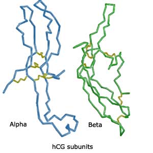
Molecular characteristics of HCG
hCG is a glycoprotein
composed of two subunits, alpha and beta, held together by ionic and
hydrophobic forces. The alpha subunit is a glycopeptide of 92 aminoacids
stabilized by five disulfide linkages. The aminoacid sequence of this
subunit is identical to that of the pituitary glycoprotein hormones,
luteinizing, follicle stimulating and thyroid stimulating hormones.
The beta subunit is a glycopeptide of 145 amino acids stabilized by six
disulfide linkages. The beta subunits of the glycoprotein hormones are
unique and give them their different biological characteristics.
Reference:
Lapthorn, A J, Harris, D.C.,
Littlejohn, A, Lustbader, J W, Canfield, R E, Machin, K J, Morgan,
F J, Isaacs, N W:
Crystal structure of human chorionic gonadotropin.
Nature, 369, 455-461, 1994
 Blastocyst
implanted in the endometrium
Blastocyst
implanted in the endometrium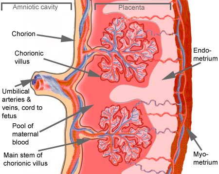 Fetal
and maternal tissues
Fetal
and maternal tissues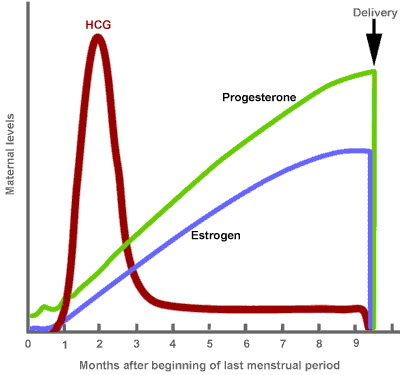 Hormone
levels
Hormone
levels