|
Exercise Physiology Laboratory |
Cardio/CNS contribution |
| |
Many factors contribute to
the changes observed during and immediately after exercise. The
following will be covered:
Cardio-CNS contribution
Respiratory contribution
Changes at the muscular level
Energy expenditure during exercise
|
|
Cardiac output may
increase to 35L/min in well-trained athletes. In untrained
individuals it can reach 20-25L/min. Most of the increase in
cardiac output goes to the exercising muscles.
There is an increase
in blood flow to skin (dissipation of heat) and to the heart
(increased work performed by the heart). Increased flows are the
result of local arteriolar vasodilation. In both skeletal and
cardiac muscles, vasodilation is mediated by local metabolic
factors, and in the skin, it is achieved mainly by a decrease in
the firing of sympathetic neurons supplying skin vessels.
Simultaneously with vasodilation in these three regions, a
vasoconstriction occurs in the kidneys and gastrointestinal
organs, due to an increase in activity of sympathetic neurons
supplying them. |
|

Distribution of the systemic cardiac
output at rest
and during strenuous exercise |
|
Vasodilation of
arterioles in the skeletal and heart muscles and skin causes a
decrease in total peripheral resistance to blood flow. This
decrease is partially offset by vasoconstriction of arterioles
in other organs. But the vasodilation in muscle arterioles is
not compensated, and the net result is a marked decrease in
total peripheral resistance to blood flow.
The mean arterial
pressure is the arithmetic product of
the cardiac output and
the total peripheral resistance (P=COxR).
During exercise, the cardiac output increases more than the
total resistance decreases, so the mean arterial pressure
usually increases by a small amount. Pulse pressure, in
contrast, markedly increases because of an increase in both
stroke volume and the speed at which the stroke volume is
ejected.
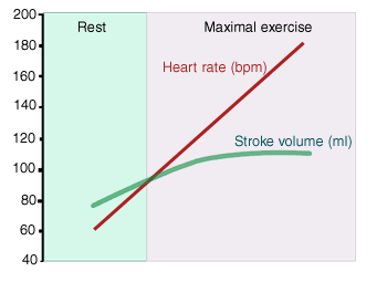
The cardiac output
increase is due to a large increase in heart rate and a small
increase in stroke volume.
|
|
 |
|
The heart rate
increases because of a decrease in parasympathetic activity of
SA node combined with increased sympathetic activity.
The stroke volume
increases because of increased ventricular contractility,
manifested by an increased ejection fraction and mediated by
sympathetic nerves to the ventricular myocardium.
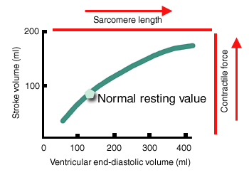
End-diastolic volume
increase slightly. Because of this increased filling, the
Frank-Starling mechanism also contributes to the increased
stroke volume (stroke volume increases when end-diastolic volume
increases).
|
|
 |
|
Cardiac output can
be increased to high levels only if the peripheral processes
favoring venous return to the heart are simultaneously activated
to the same degree. Factor promoting venous return:
-
increased activity
of the skeletal-muscle pump.
-
increased depth and frequency
of respiration; respiratory pump.
-
sympathetically
mediated increase in venous tone
-
greater ease of
blood flow from arteries to veins.
Control of
sympathetic outflow
One or more discrete control centers in the brain are activated
by output from the cerebral cortex. Descending pathways from
these centers transmit these centers’ activity to the
appropriate autonomic preganglionic neurons eliciting the firing
patterns typical for exercise. These centers become activated
before the exercise started.
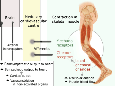
Once exercise
is started, local chemical changes in the muscle can develop,
particularly during high levels of exercise, because of
imperfect matching between blood flow and metabolic demands.
These changes activate chemoreceptors in the muscle. Afferent
input from these receptors goes to the medullary cardiovascular
centers. The result is a further
increase in heart rate, myocardial contractility, and
vasoconstriction in the nonactivated organs. Mechanoreceptors of
the exercising muscle are also stimulated and provide an
excitatory input to the medullary cardiovascular center.
|
|
 |
|
The arterial
baroreceptors
As mean and
pulsatile pressure increase, baroreceptors should respond to
increase parasympathetic and decrease sympathetic outflows, a
pattern designed to counter the rise
in arterial pressure. During exercise the
exact opposite occurs:
the arterial baroreceptors increase the arterial pressure during
exercise. The reason is that one of neuronal component of the
central command output goes to the arterial baroreceptors and
‘resets’ them upwards as exercise begins. The resetting causes a
decrease firing frequency in the baroreceptors, signalling for
decreased parasympathetic and increase in sympathetic outflow. |
|
|
|
To continue with the next section:
respiratory contribution, click here |