|
Cardiovascular Laboratory |
ECG> Introduction |
| |
The electrocardiograph (abbreviated ECG or EKG) is a
device that picks up electrical activity originating in the heart from the surface of the
body. In the clinic, the ECG is one of the most commonly used diagnostic machines.
The recording produced by the electrocardiograph is called an electrocardiogram,
also abbreviated ECG or EKG. |
|
Electrocardiogram
Waveform |
|
|
|
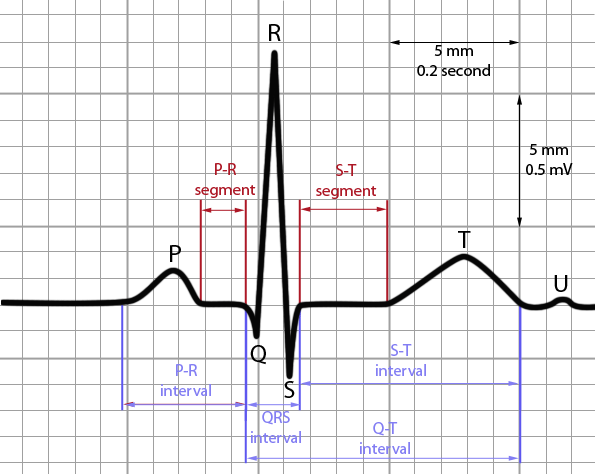
| The figure to the right shows a typical ECG.
Three characteristic features of the waveform are easily identified: the P wave,
the QRS complex, and the T wave. The P wave is associated with the activation of the
atria, the QRS complex with the activation of the ventricles, and the T wave with
repolarization of the ventricles. |
 |
|
Electrocardiogram
Intervals |
|
|
|
- The P-R interval is the time from the beginning of the P wave
to the start of the QRS complex.
- The QRS interval or duration or
width is the time from the beginning to
the end of the QRS complex.
- The QT interval is the time from the beginning of the QRS
complex to the end of the T wave.
- The RR interval is the time from the peak of one R wave to
that of the following R wave.
|
 |
|
Electrocardiograph:
Technical Principles |
| The electrocardiograph is
essentially an electronic device that amplifies the very small potentials present at the
surface of the body, so that they can be displayed on a video screen or recorded
permanently on paper. The signal is picked up by electrodes placed at certain well-defined
anatomical positions on the body surface. |
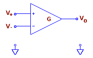
The heart of the
ECG is an electronic amplifier with two input terminals, a non-inverting input terminal
(+) and an inverting input terminal (-). The output voltage Vo is
simply proportional to the difference between the voltages V+ and V-
appearing at the two input terminals: |
 |
| Vo
= G( V+ - V- ), |
where G is the gain
of the amplifier. The amplifier is thus termed a differential amplifier, since it
measures the difference between two voltages. |
| You will remember from
your basic physics courses that electrical voltage or potential, unlike length or mass, is
not an absolute quantity, but rather a relative quantity, in that potential itself cannot
be measured, only differences in potential. Thus V+ and V-
are each measured with respect to some third reference point that is arbitrarily taken to
be at zero potential. In electrocardiography, this point is the right leg. |
| The differential
amplifier has the advantage that any component of the signal appearing simultaneously at
both inputs is cancelled out and so does not appear at the output. This
"common-mode
rejection" is important, since electrical 115-volt power wiring in a building can induce
signals at 60 Hz (the power line frequency) on the body surface that are many times larger
than the ECG signal itself. Use of a differential amplifier prevents this large
spurious signal from swamping out the ECG signal. |
|
|
To continue with the next section:
ECG Setup, click here |