|
Cardiovascular Laboratory |
ECG> Experiments |
| |
The ECG trace depends on the lead
configuration, which sets the orientation of the lead axis relative to the heart.
The
standard limb leads have the disadvantages that the three electrodes are all in the
same plane (frontal plane) of the body, so that one is only recording a
projection of the three-dimensional spread of depolarization and
repolarization in that plane. |
|
|
|
1)
Inspecting the ECG |
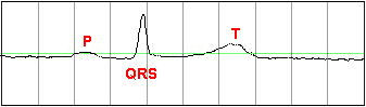
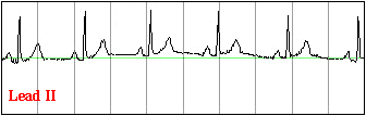
- While displaying Lead II, the P-wave, the QRS-complex, and
the T-wave are identified. In the chart program, experiment with different settings for
the time scale and the voltage scale so that, e.g. the waveform
is not too big or small in amplitude.
|
|
|
|
|
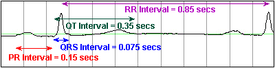
2)
Identifying Waves and Intervals |
- The time scale is set to an appropriate value for
displaying the overall shape of the P-QRS-T waveform. The various complexes and
intervals are examined. The RR, PR, QRS and QT intervals are measured, and compared
to the normal ranges given in the table to the right (in seconds).
|
| Interval |
Min |
Max |
| RR |
0.6 |
1.2 |
| PR |
0.12 |
0.20 |
| QRS |
|
0.10 |
| QT |
|
0.42 |
|
|
|
|
3)
Effect of Lead Placement |
- The white (Neg) electrode on the
right arm is moved from its position on the wrist to a
new position somewhere above the elbow.
|
 |
 For convenience, the
connections of the ECG electrodes are usually made at the ends of the limbs: at the wrists
and ankles. However, since the limbs act as conductors, they can be viewed as an
extension of the patient cable lead, and so it makes no difference where the electrodes
are placed along the limb length. For convenience, the
connections of the ECG electrodes are usually made at the ends of the limbs: at the wrists
and ankles. However, since the limbs act as conductors, they can be viewed as an
extension of the patient cable lead, and so it makes no difference where the electrodes
are placed along the limb length.
|
- After returning the white electrode to its original position, the
subject extends the right arm outwards and holds it horizontally in mid-air away from the
body.
|
 |
|
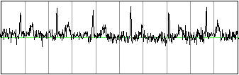
The above ECG trace appears
very noisy, because the recording is also picking up the EMG activity from the muscles
used in extending the arm outwards. |
|
|
4)
Effect of Respiration |
- The subject takes a deep slow breath, and then exhales slowly
(inhaling for 5 seconds, and exhaling for five seconds).
|
 |
|
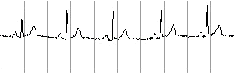
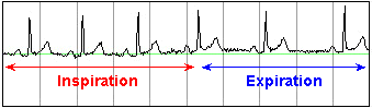
In
sinus arrhythmia, the heart rate varies with the
phase of respiration. The heart rate typically increases during inspiration and
decreases during expiration. Therefore, as observed, the R-R interval is longer
during expiration. These changes are mediated through vagal reflexes. Sinus
arrhythmia is more common in young healthy athletes. |
|
|
5)
The Timing of the Heart Sounds |
- One member of the group listens with the stethoscope to the
subject's heartbeat to determine where the two well-separated heart sounds fall on the ECG
trace.
|
 |
| |
|
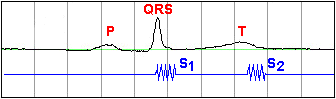
The first heart sound
S1 is due to the closure of the mitral and
tricuspid valves at the start of ventricular systole. The second heart sound
S2 is due
to the closure of the aortic and pulmonary valves. Click here for more on the heart valves |
|
|
6)
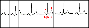
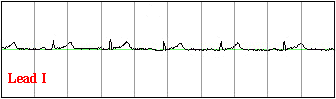
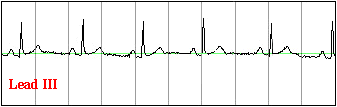
Changes in Morphology with Leads |
- The cables are connected so as to
record from lead I. The group
should describe the changes seen with respect to lead II, and attempt to explain them.
|
- The cables are connected so as to
record from lead III. The group
should describe the changes seen with respect to lead II, and attempt to explain them.
|
|
|
 |
 |
 |
|
Recall that the R wave is due
to the activation (depolarization) of the major portion of the ventricles. From the
sample data above, it is evident that the lead whose axis is
most parallel to the
direction of the subject's ventricular depolarization is lead II. (The R wave is largest
in lead II.) The R wave is very small in lead I. We can therefore conclude
that for this subject the direction of ventricular depolarization is more close to being
perpendicular
to lead I. |
|
|
To continue with the next section:
ECG Disorders, click here |