|
Biological Signals Acquisition |
EOG tests > intro |
| |
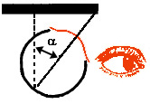
Recording eye movements of a
stationary subject: a reference electrode is placed on the forehead,
electrodes are placed on the right and left temples for lateral eye
movement detection (for vertical eye movement detection, one can also
place electrodes above and below an eye). This technique is also an
indirect way to evaluate the tracking and scanning of visual targets. |
| |
|
| |
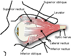
| We move our eyes
constantly during our daily activities to keep
our line of sight pointed at a target of
interest. In order to generate an eye movement
along any axis, there are three antagonistic
pairs of muscles which are attached to the globe
of the eye. |
| These
sets of muscles function to move the eye
horizontally (left versus right), vertically (up
versus down) and torsionally (clockwise versus
counter clockwise). There are four different types of
conjugate eye movements. These eye movements fall
into two specific categories:
- Eye movements
that function to stabilize the position
of the eye in space during head movements
(Reflex eye movements).
- Eye movements
that function to redirect the line of
sight to follow a moving target or to
attend to a new target of interest
(Voluntary eye movements).
|
 |
|
The Technique: Electro-oculography (EOG) |
 |
In
the 1920's, it was discovered that by placing
electrodes on the skin in the region of the eyes,
one could record electrical activity which
changed in synchrony with movements of the eye in
the head. It was initially believed that these
potentials reflected the action potentials in the
muscles that are responsible for moving the eyes
in the orbit. |
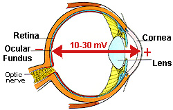
| However, it is now generally agreed that these
electrical potentials are generated by the
permanent potential difference which exists
between the cornea and the ocular fundus
(cornea-retinal potential, 10-30mV: the cornea
being positive). |
| This
potential difference sets up an electrical field
in the tissues surrounding the eye. As the eye
rotates, the field vector rotates
correspondingly. Therefore, eye movements can be
detected by placing electrodes on the skin in the
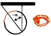
area of the head around the eyes. Vertical
movements of the eyes are best measured by
placing the electrodes on the lids, while
horizontal eye movements can be best measured by
placing the electrodes on the external canthi (the
bone on the side of the eye). |
|
Limitations
of the Technique |
|
The
underlying assumption of this method of recording
eye movements is that the movement of the
electric field in the conducting tissues
surrounding the eye is related, in a simple
(usually assumed to be linear) way to the
movements of the eye itself. Due to the
non-uniformity of these tissues and the shapes of
the tissues surrounding them, this can only be an
approximation to the biological reality. However,
for horizontal eye movements within the range of
30 degrees, the potential measured is assumed to
be linear to the actual movement of the eye in
the orbit. The resolution of EOG is considered to
be about 1 degree. Because it is a relatively
simple technique, EOG is still commonly used
clinically for testing eye movements in patients. |
For a
fixed eye position, the EOG is far from being
constant in magnitude, but can be influenced by a
number of external factors. These factors include
- the noise
generated between the electrodes'
contacts and the skin
- the metabolic
state of the tissues (pO2, pCO2, and temperature)
- visual
stimulation
- contraction
of facial muscles
In addition,
recorded EOG, particularly for vertical eye
movements, is quite sensitive to movements of the
eye lids. In summary there are a number of
external factors which can complicate the
interpretation of the EOG, and for that reason
EOG is considered highly sensitive to artifacts.
The considerable artifacts which can be
introduced through the contact between the
electrode contacts and the skin can be minimized
by reducing the resistance between the electrodes
and the skin.
|
 |
 |
 |
|
To continue to the next section, EOG- procedure,
click here |