|
Blood
Laboratory |
Blood cell indices
>
RBC count |
| |
Among the most common hematological tests performed are
those which determine the red blood cell count, the hematocrit, the hemoglobin content,
ABO/Rh Blood Group typing, and the total and differential white blood cell count. Each one
of these measurements may be carried out on a single drop of blood. |
|
Review the topics in your textbook. |
|
Theory |
|
A convenient and inexpensive way to
count blood cells is through the use of the hemocytometer. This instrument is a special
microscope slide on which precise grids have been etched within a counting chamber
designed to hold an exact volume of diluted blood sample. The blood cell determination
involves counting cells in several squares of the grid and obtaining an average number.
That number is multiplied by a factor that compensates for the amount of dilution. The
final result expresses the number of red blood cells per cubic millimeter of the original
blood sample.
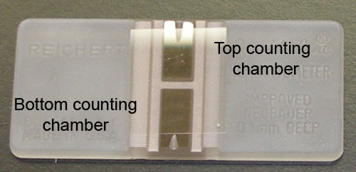
The picture above shows a hemocytometer,
on top of which a coverslip (thin square glass slide) was placed to
delimit the area of each of the counting chambers, top and bottom.
The groove (present on some hemocytometer models) on the edge of
each counting chamber facilitates the positioning of the pipette tip
and the loading of the diluted blood sample.
|
|
Procedure |
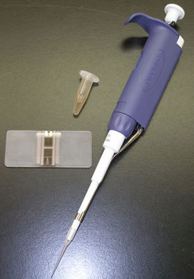
The picture above shows a microcentrifuge
tube (sometimes referred to as an eppendorf tube) containing the diluted
blood sample in Gower's solution, a 20
ml
adjustable pipette (or pipetman) and the hemocytometer. |
In order to be able to effectively count cells, venous blood in EDTA (as anticoagulant)
must be added to an
isotonic solution (Gower's solution) which prevents lysis and crenation
of erythrocytes. Gower's solution also contains a fixative which acts as
a preservative and prevents agglutination of cells if counting cannot be
done within an hour.Preparation of the diluted blood sample:
Volume to volume dilution is one in which a unit volume of a solute
is combined with an appropriate volume of a solvent liquid to achieve
the desired concentration. The dilution factor is the total number of
unit volumes in which the solute is dissolved in the solvent. This
dilution factor method is based on ratios.
In this exercise, it is desirable to dilute blood 1:200; one "unit" of
blood is to be diluted into 199 units of Gower's solution, for a
dilution factor of 200. How much blood should you add to the Gower's
solution to get a final volume of 1ml?
Loading the hemocytometer:
After adequate
mixing, the diluted blood is introduced into the counting chamber where
the cell count is performed microscopically.
|
|
Caution:
|
The 20 ml
pipette tip is placed on the hemocytometer at the edge of the coverslip.
The plunger of the adjustable pipette is slowly pressed so that the
sample flows between the hemocytometer's raised shiny surface and the
coverslip. Stop when the sample touches the three sides of the chamber
(about 10 ml);
do not overflow into the moat which surrounds the chambers. |
|
Counting |
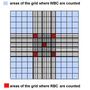
| Observe the grid of the hemocytometer
below. Different areas are used for counting red blood cells and white blood cells. Red
cells are counted in the areas indicated in red. |
Consider the following: The central
grid of 25 squares is 1mm x 1mm in area and 0.10 mm deep, and holds 0.1ml
of liquid.
The dilution factor is 1:200.
Convert the number of red blood cells that are counted in 5 squares to the number of red
blood cells/µl. (N.B. 1 µl
(microliter) = 1 cubic mm ).
Since only 5 out of the 25 squares (1:5)
are counted, you must multiply your count by 5.
Since you have diluted your sample by 200 (1:200), you must also
multiply your count by 200 to arrive at the original concentration in
the body.
You obtain a count which is contained in a volume of 0.1µl (25 squares).
So you must also multiply by 10 to express the count per cubic mm. |
 |
Click here to open a window which mimics
what you might see looking through the 40x objective of a microscope.
Only the center of the grid shown above is visible (where the 5 squares
are coloured red). Try to verify this by observing the fineness of the
grid as you move to the edges of the visible field. Count the red cells
in the five squares and determine the red cell count as described above.
Important note: To avoid counting the same cells twice, cells that
are touching the lines at the tops and left sides of the squares are counted, but cells
that are touching the bottoms and right sides of the squares are not counted.
| Female: |
3.9-5.6 million/µl |
| Male: |
4.5-6.5 million/µl |
|
|
To
continue with the next section, Hematocrit, click here |