|
Biomedical Signals Acquisition |
EEG > introduction |
| |
Recording the electrical
activity of the brain from the scalp: an
introduction to the acquisition of biological signals |
| |
|
| |
The
electroencephalogram (EEG) is a recording of the electrical activity of
the brain from the scalp. The recorded waveforms reflect the cortical
electrical activity.
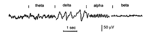
Signal intensity: EEG activity is quite small, measured in microvolts (mV).
Signal frequency: the main frequencies of the human EEG waves are:
- Delta:
has a frequency of 3 Hz or below. It tends to be the highest in
amplitude and the slowest waves. It is normal as the dominant rhythm
in infants up to one year and in stages 3 and 4 of sleep. It may
occur focally with subcortical lesions and in general distribution
with diffuse lesions, metabolic encephalopathy hydrocephalus or deep
midline lesions. It is usually most prominent frontally in adults
(e.g. FIRDA - Frontal Intermittent Rhythmic Delta) and posteriorly
in children e.g. OIRDA - Occipital Intermittent Rhythmic Delta).
- Theta:
has a frequency of 3.5 to 7.5 Hz and is classified as "slow"
activity. It is perfectly normal in children up to 13 years and in
sleep but abnormal in awake adults. It can be seen as a
manifestation of focal subcortical lesions; it can also be seen in
generalized distribution in diffuse disorders such as metabolic
encephalopathy or some instances of hydrocephalus.
- Alpha:
has a frequency between 7.5 and 13 Hz. Is usually best seen in the
posterior regions of the head on each side, being higher in
amplitude on the dominant side. It appears when closing the eyes and
relaxing, and disappears when opening the eyes or alerting by any
mechanism (thinking, calculating). It is the major rhythm seen in
normal relaxed adults. It is present during most of life especially
after the thirteenth year.
- Beta:
beta activity is "fast" activity. It has a frequency of 14 and
greater Hz. It is usually seen on both sides in symmetrical
distribution and is most evident frontally. It is accentuated by
sedative-hypnotic drugs especially the benzodiazepines and the
barbiturates. It may be absent or reduced in areas of cortical
damage. It is generally regarded as a normal rhythm. It is the
dominant rhythm in patients who are alert or anxious or have their
eyes open.
|
|
 |
| Variables used in the classification
of EEG activity |
|
|
|
Frequency refers
to rhythmic repetitive activity (in Hz). The frequency of EEG activity
can have different properties including:
- Rhythmic. EEG activity
consisting in waves of approximately constant frequency.
- Arrhythmic. EEG activity in
which no stable rhythms are present.
- Dysrhythmic. Rhythms and/or
patterns of EEG activity that characteristically appear in patient
groups or rarely or seen in healthy subjects.
|
|
|
|
Voltage refers to the
average voltage or peak voltage of EEG activity. Values are dependent,
in part, on the recording technique. Descriptive terms associated with
EEG voltage include: |
-
Attenuation
(synonyms: suppression, depression). Reduction of amplitude
of EEG activity resulting from decreased voltage. When
activity is attenuated by stimulation, it is said to have
been "blocked" or to show "blocking".
- Hypersynchrony. Seen
as an increase in voltage and regularity of rhythmic
activity, or within the alpha, beta, or theta range. The
term implies an increase in the number of neural elements
contributing to the rhythm. (Note: term is used in
interpretative sense but as a descriptor of change in the
EEG).
- Paroxysmal. Activity
that emerges from background with a rapid onset, reaching
(usually) quite high voltage and ending with an abrupt
return to lower voltage activity. Though the term does not
directly imply abnormality, much abnormal activity is
paroxysmal.
|
|
|
|
|
Morphology refers to the
shape of the waveform. The shape of a wave or an EEG pattern is
determined by the frequencies that combine to make up the waveform and
by their phase and voltage relationships. Wave patterns can be described
as being:
-
Monomorphic.
Distinct EEG activity appearing to be composed of one dominant
activity
-
Polymorphic.
distinct EEG activity composed of multiple frequencies that combine
to form a complex waveform.
-
Sinusoidal. Waves
resembling sine waves. Monomorphic activity usually is sinusoidal.
-
Transient. An
isolated wave or pattern that is distinctly different from
background activity.
| a) Spike: a transient with a
pointed peak and a duration from 20 to under 70 msec. |
| b) Sharp wave: a transient
with a pointed peak and duration of 70-200 msec. |
|
|
|
|
Synchrony refers to the simultaneous appearance of rhythmic or
morphologically distinct patterns over different regions of the head,
either on the same side (unilateral) or both sides (bilateral). |
|
|
|
Periodicity refers to the distribution of patterns or elements in time
(e.g., the appearance of a particular EEG activity at more or less
regular intervals). The activity may be generalized, focal or
lateralized. |
|
|
|
|
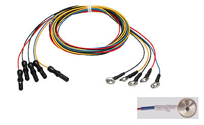
| Small metal
discs usually made of stainless steel, tin, gold or silver covered
with a silver chloride coating. They are placed on the scalp in
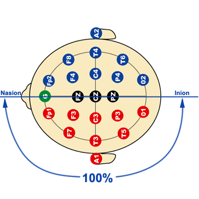
special positions. These positions are specified using the International
10/20 system. Each electrode site is labeled with a letter and a number.
The letter refers to the area of brain underlying the electrode e.g. F-
Frontal lobe and T - Temporal lobe. Even numbers denote the right side
of the head and odd numbers the left side of the head. |
Copyright
ADInstruments. All rights reserved. |
EEG cables showing the disc
electrodes to which electrode gel is applied and applied to the
subject's scalp. |
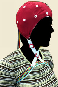
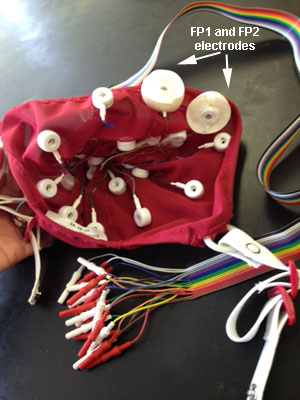
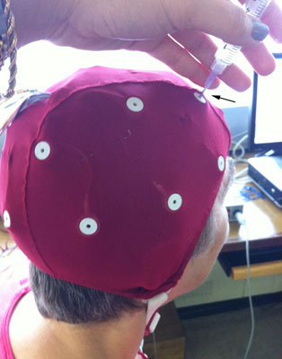
Many recording systems use a cap into
which electrodes are embedded; this facilitates recordings when high
density arrays of electrodes are needed or when comparing recording
sites. The image to the right shows the inside of such a cap. |
 |
|
|
It acts as a
malleable extension of the electrode, so that the movement of the
electrodes cables is less likely to produce artifacts. The gel maximizes
skin contact and allows for a low-resistance recording through the skin.
The electrolytic gel is injected into each cavity until a small
amount comes out the hole in the mount. With a moderate amount of
downward pressure,the syringe with a blunt needle is rapidly rocked back
and forth. |
|
|
| A measure of the
impediment to the flow of alternating current, measured in ohms at a
given frequency. Larger numbers mean higher resistance to current flow.
The higher the impedance of the electrode, the smaller the amplitude of
the EEG signal. In EEG studies, should be at lest 100 ohms or less and
no more than 5 kohm. |
| Electrode
positioning (10/20 system) |
|
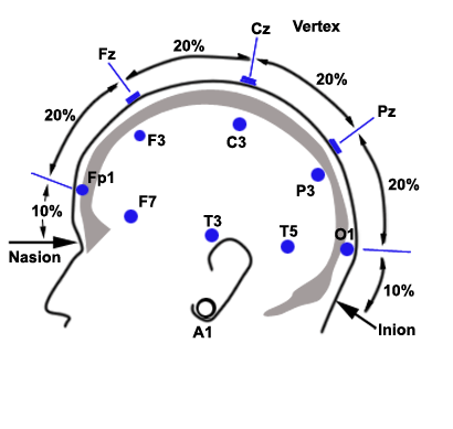
| The standardized
placement of scalp electrodes for a classical EEG recording has become
common since the adoption of the 10/20 system. The essence of this
system is the distance in percentages of the 10/20 range between
Nasion-Inion and fixed points. These points are marked as the Frontal
pole (Fp), Central (C), Parietal (P), occipital (O), and Temporal (T).
The midline electrodes are marked with a subscript z, which stands for
zero. The odd numbers are used as subscript for points over the left
hemisphere, and even numbers over the right. |
 |
 |
|
10/20 System of electrode placement |
|
|
|
Montage means the placement of the electrodes. The EEG can be monitored
with either a bipolar montage or a referential one. Bipolar means that
you have two electrodes per one channel, so you have a reference
electrode for each channel. The referential montage means that you have
a common reference electrode for all the channels. |
|
|
| Artifacts |
| The
biggest challenge with monitoring EEG is artifact recognition and
elimination. There are patient related artifacts (e.g. movement,
sweating, ECG, eye movements) and technical artifacts (50/60 Hz
artifact, cable movements, electrode paste-related), which have to be
handled differently. There are some tools for finding the artifacts. For
example, FEMG and impedance measurements can be used for indicating
contaminated signal. By looking at different parameters on a monitor,
other interference may be found. |
| |
|
|
Electrodes used in EEG recording do not
discriminate the electrical signals they receive. The recorded activity
which is not of cerebral origin is termed artifact and can be divided
into physiologic (generated from the subject from sources other than the
brain) and extraphysiologic artifacts arise from outside the body
(equipment including the electrodes and the environment). |
|
Electromyogram (EMG)
activity |
|
EMG activity are common artifacts: the
myogenic potentials generated in the frontalis muscles (raising
eyebrows) and the temporalis muscles (clenching of jaw muscles) are of
shorter duration than those generated in the brain. These artifacts can
be identified on the basis of duration, morphology and rate of firing
(frequency). Particular patterns of EMG artifacts can occur in some
movement disorders: essential tremor and Parkinson disease can produce
rhythmic 4 to 6 Hz sinusoidal waveforms. |
|
Eye movements |
|
The eyeball acts as a dipole with a positive
pole oriented anteriorly (cornea) and a negative pole oriented
posteriorly (retina). When the globe rotates about its axis, it
generates a large amplitude alternate current field detectable by any of
the electrodes positioned near the eye. A blink causes the positive pole
(the cornea) to move closer to frontopolar FP1, FP2 electrodes,
producing symmetric downward deflections. |
 |
|
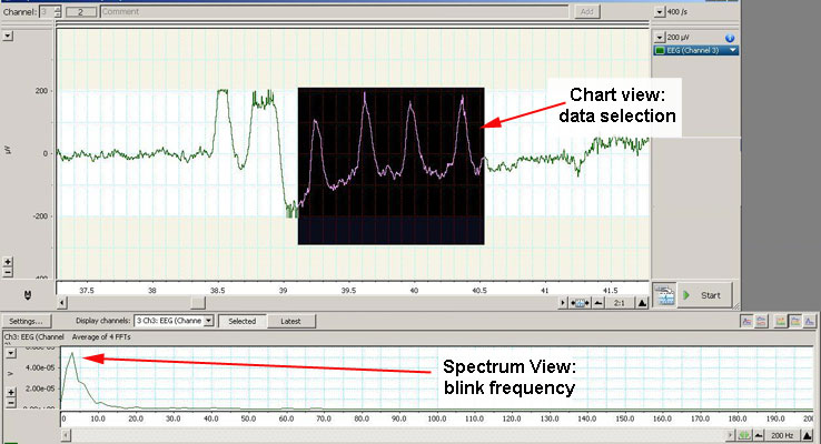
In the above example, the subject was blinking while the chart view and
the recording was active (notice the four higher amplitude waves). The
spectrum view window calculated and displayed a dominant frequency of 3
Hz which was the blinking frequency. |
|
Skin artifacts |
|
A further difficulty arises due to
properties of certain layers of the skin. A significant DC potential
exists between the stratum corneum and the stratum granulosum and any
local deformation of the skin will alter this potential. The only
reliable way to eliminate the source of artifact is to to create a low
resistance pathway through the layers of skin by skin cleaning (alcohol
swab). Also, sodium chloride (electrolyte) from sweating reacting with
metals of the electrodes may produce a slow baseline drift. |
|
Electrodes |
|
Surface electrodes such as
the ones used in EEG must create an interface between an ionic solution
(the subject) and a metallic conductor (the electrode). This leads to a
half-cell potential which can be quite large relative to the signal
being recorded. To minimize this problem of polarization of the
electrode, some electrodes are coated with silver chloride, but all are
maintained away from the skin through an intermediate layer of
conductive paste. Touching the electrodes during recording can produce
artifacts. An electrode which is not contacting the skin very well acts
like an antenna with resulting 60-cycle interference (see recording
below). |
|
60-Hz artifact |
| The
problem arises when the impedance of one of the active electrodes
becomes significantly large between the electrodes and the ground of the
amplifier. In this situation, the ground becomes an electrode that,
depending on its location, produces the 60-Hz artifact. Interference
from high-frequency radiation from other electronic devices can overload
EEG amplifiers. |
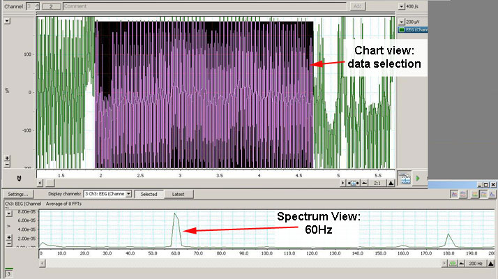 |
| In the above recording,
there was a very poor contact of the electrodes with the scalp of the
subject; the spectrum view shows a dominant frequency of 60 Hz. |
|
|
| It
is the key to electrophysiological equipment. It magnifies the
difference between two inputs. An unwanted signal that is common to the
two inputs will be subtracted. |
|
|
| The
standard filtering settings for routine EEG are:
Low frequency filter: 1 Hz
High frequency filter: 50-70 Hz |
|
Click here to continue with the alpha waves experiments |
| |
| |