In a restrictive lung disease,
the compliance of the
lung is reduced, which increases the stiffness of the lung and
limits expansion. In these cases, a greater pressure (P)
than normal is required to give the same increase in volume (V).
Common causes of decreased lung compliance are pulmonary fibrosis,
pneumonia and pulmonary edema.
In
an obstructive lung disease, airway obstruction causes
anincrease in resistance.
During normal breathing, the pressure volume relationship is no
different from in a normal lung. However, when breathing rapidly,
greater pressure is needed to overcome the resistance to flow, and the
volume of each breath gets smaller. Common obstructive diseases include
asthma, bronchitis, and emphysema.
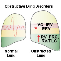
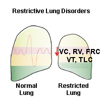
Changes in Lung Volumes
Observe the changes in lung volumes from
normal for restrictive and obstructive lung disorders. In the obstructed
lung, respiration ends prematurely, thus increasing RV and FRC. In the
restricted lung, volumes are small because inspiration is limited due to
reduced compliance.
The FVC test allows one to
clearly distinguish between the two disease types. Notice in the
obstructed lung (below left), how FVC is smaller than normal, but also
that FEV1
is much smaller
than normal. This is because it is very difficult for a person with an
obstructive disease (eg. asthma) to exhale quickly due to the increase
in airway resistance. As a result, the FEV1/FVC
ratio will be much lower than normal, for example 40% as opposed to 80%.
In the restricted lung, the
FVC is again smaller than normal, but the FEV1
is relatively large in comparison. i.e. the FEV1/FVC
ratio can be higher than normal, for example 90% as opposed to 80%. This
is because it is easy for a person with a restricted lung (e.g fibrosis)
to breathe out quickly, because of the high elastic recoil of the
stiff lungs.