|
Compound Action Potential |
Experimental preparation |
| |
In order to record a CAP
successfully, one needs to take special care to dissect out the longest
nerve possible and to keep it moist at all times. It is also important
to ensure the nerve makes good contact with the stimulating and the
recording electrodes in the bath. |
|
|
|
Dissecting out the Frog
Sciatic Nerve |
|
|
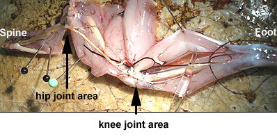
The schematic view of the frog sciatic nerve to
the right shows the path of the nerve from the spinal insertion points down to the ankle. The frog sciatic nerve dissection is a delicate operation.
During the dissection, the nerve must not be touched with the fingers, cut muscle, frog
skin, or with any metal instruments. Care must be taken to keep the preparation moist at
all times, and to not stretch the nerve unduly. |
|
|
Glass probes are used to dissect out the frog
sciatic nerve, by gently separating the nerve from the surrounding muscle masses. Great
care must be taken when freeing the nerve from tendons at the hip and knee joints. It is
desirable to obtain as long a length of nerve as possible, from the vertebral column down
to the foot. |
|
 |
|
A small slip of bone/muscle is left attached
to the nerve at the proximal end. A piece of thread is tied to the nerve at the
distal
end. |
|
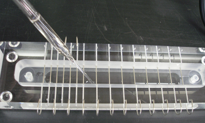
The lower reservoir of the
nerve bath (above) is filled with frog
Ringer's solution, making sure that the Ringer's solution is not touching any of the
wires. The nerve is placed in the chamber with the bone end near the two stimulating
electrodes (anode and cathode). The lid of the chamber is then
placed firmly on top to preserve the humidified air in the bath. |
|
 |
|
|
|
|
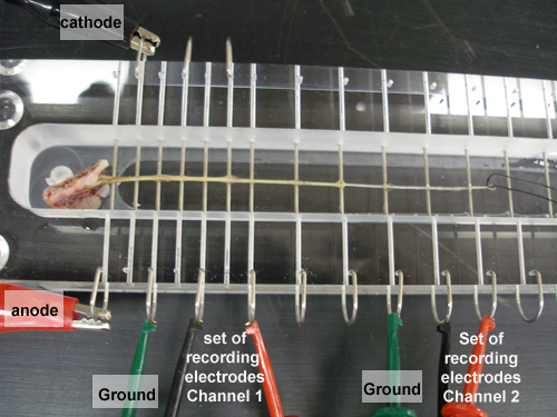
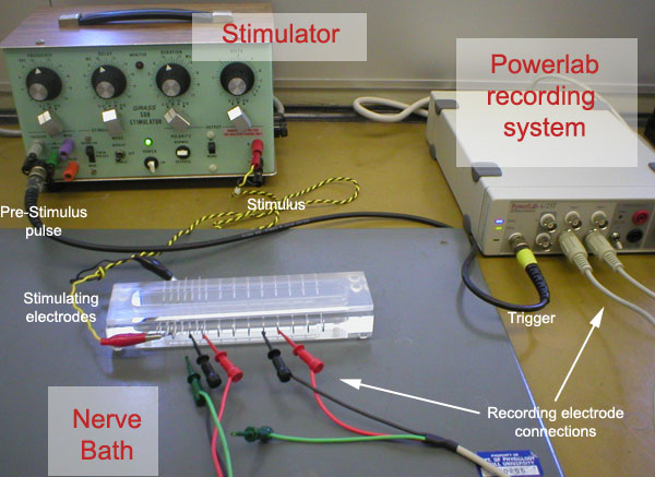
Observe the following setup: |
|
 |
|
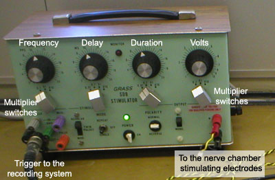
The Grass stimulator provides two output pulses. One set
of leads, from the stimulator to the pair of stimulating electrodes, carries the Stimulus
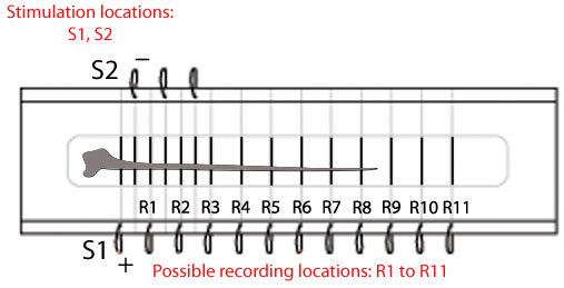
Pulse that will activate the nerve. Because the
CAP is initiated at or near the cathode, and the anodal action slows
down or may even suppress the CAP, the cathode should always be closer
to the recording electrodes. The nerve will be stimulated repeatedly, and the
Amplitude, Duration and Frequency knobs and switches control the corresponding parameters
of the Stimulus Pulse. (The value of each parameter is the product of the dial readings on
the corresponding range switch and dial.) |
|
A second lead from the stimulator (a
two-conductor, co-axial cable) carries a Pre-stimulus Trigger Pulse to the
Trigger connector of the Powerlab recording system. This narrow Trigger Pulse
precedes the Stimulus Pulse and is interpreted by the Analogue I/O board
in "Powerlab" as a signal to start sampling a new sweep of data. |
 |
|
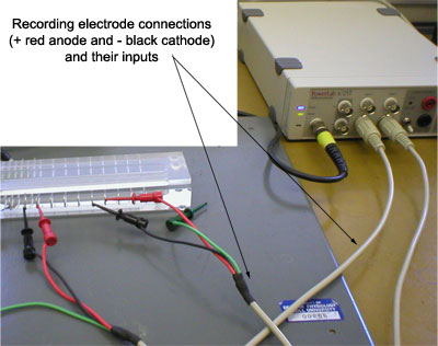
Satisfactory recording of the electrical
response of the nerve depends on a pair of shielded cables leading from the
two pairs of Recording
Electrodes to the input connectors (CH1 and CH2) of the recording system. The shields of these coaxial cables reduce electrical noise.
|
 |
|
The stainless steel
electrodes in the bath are equally spaced. Connection to these wires
depend on the length of the dissecting nerve. Conduction velocities and
refractory measurements are facilitated by the fact that one can record
from different sites along the nerve. |
|
Modified from ADinstruments.
All Rights Reserved.
|
|
Click here to continue
with the topic of the Characteristics of the CAP |